Brain imaging identifies different types of depression

Biological markers could enable tailored therapies that target individual differences in the symptoms of depression (Scientific American, 2017).
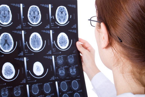
New research, published in Nature Medicine, by Conor Liston, a neuroscientist and psychiatrist at Weill Cornell Medical College, and his colleagues seems to point toward a long-sought goal in psychiatry, biological markers to distinguish different kinds of depression. The researchers used a noninvasive technique called functional magnetic resonance imaging (fMRI) to measure the strength of connections between neural circuits in the brain. Analysing fMRI scans from more than a thousand people, of whom about 40% had been diagnosed as depressive, the team identified four subtypes of depression. If confirmed in additional studies, the findings could enable clearer diagnoses and pave the way for personalised therapies targeting brain networks found to be awry in individual patients.
The study, published in December, grew out of an observation Conor Liston made as an MD/PhD student. He had conducted experiments in rats and found that stress reduced neural connections in a brain area called the prefrontal cortex, which controls mental flexibility, the ability to adapt thinking to new situations and to overcome habitual responses. Conor Liston also studied stress in students preparing for their medical licensing exams. Like the rats, stressed students displayed abnormal electrical activity in brain circuits that involve mental flexibility. Luckily, getting a month off after the test allowed their faulty networks to recover, suggesting the brain is more resilient than expected. In a later study that Conor Liston conducted with Weill Cornell psychiatrist Marc Dubin, the brain imaging researchers detected similar network changes in people who are depressed, but only in a small subset of these patients. That intrigued Conor Liston. It seemed to him that stress, or something like it, throws off the flexibility circuits in certain depressed individuals, whereas other people become depressed for different reasons. He said that would be consistent with the view that depression “is not just one biological thing.”
To look for new biomarkers, the Weill Cornell team used a method called resting-state fMRI to check for differences in brain connectivity between depressed and healthy people. The procedure scans the brain while a person lies on a bed for five minutes. The resulting data is complex and messy. Brain fMRI measurements are sensitive to minuscule differences between subjects such as whether people look around the room or shut their eyes during the scan. To do a rigorous analysis, Conor Liston knew he needed a mountain of data, far more than he could collect on his own. He said “I went around and begged a lot of people I knew, and some I didn’t know, who had collected data the same way we did.” He ended up with brain scans from 1,188 individuals, some healthy and some depressed, studied at seventeen research sites worldwide. He said having that much data yielded enough statistical power that “we didn’t have to constrain ourselves to [analysing] just a few regions” of the brain. For each subject the team examined two hundred and fifty eight brain areas, measuring how strongly each connects with other areas.
Using machine learning, the analysis showed depressed people could be distinguished from healthy controls based on brain connectivity differences measured by fMRI in the limbic and frontostriatal areas. The limbic system controls emotions and frontostriatal networks help coordinate motor and cognitive functions. One brain area, called the subgenual cingulate cortex, has unusually strong connections with other regions of the brain in people who are depressed. Prior imaging studies had implicated these areas in depression, and some of those analyses suggested connectivity measures could differentiate between depressed and healthy people. But the Weill Cornell team is believed to be the first to confirm findings in a separate population, an additional analysis that is seen as a mark of scientific rigour.
Nunzio Pomara, a professor of psychiatry and pathology at New York University School of Medicine who was not involved in Conor Liston’s work, said “This represents an exciting approach. It sets the stage for future studies.” He noted, though, that brain connectivity data does not address the underlying biology of depression. It does not explain what is going on at the level of cells and chemical messengers, the sorts of discoveries that guide development of new drugs. Still, he said the new fMRI analysis “goes beyond what has been done with similar neuroimaging techniques” by identifying four kinds of depressed patients on the basis of connectivity problems. Most imaging analyses merely distinguished healthy and depressed people.
In the new study, the fMRI-based subdivisions could be linked to particular symptoms. Patients falling into the first two subtypes reported more fatigue whereas those in the other two reported more trouble feeling pleasure. This subtyping has implications not just for diagnosis but potentially for non-pharmaceutical treatment. Relative to groups two and four, people with depression subtype one were three times as likely benefit from transcranial magnetic stimulation (TMS). This technology uses a magnet to produce small electric currents in brain areas affected by depression. Although the procedure is gaining popularity, it is generally reserved for patients who have not responded to conventional antidepressants. Marc Dubin believes in the next five years TMS be tuned to treat patients with different depression subtypes. After scanning a patient’s brain with fMRI, as done in the recent study, a physician could adjust the TMS magnet so it aims directly at the brain areas with abnormal connectivity in that person.


