Large Hadron Collider technology to help treat cancer
Technology from the ATLAS experiment at the Large Hadron Collider, where the Higgs Boson was discovered, will be used in hospitals to improve cancer treatments that employ proton beam therapy.
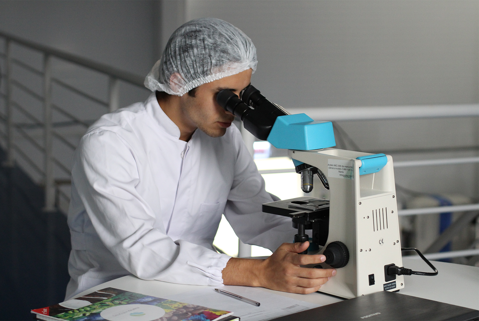
The University of Birmingham reports their researchers, who designed and built detectors for ATLAS, are using their knowhow from the Large Hadron Collider to create a new way of helping image and treat cancers.
The Large Hadron Collider is the world’s largest and most powerful particle accelerator. Beams of protons collide at the centre of the ATLAS detector creating new particles from the debris at the collision point.
Now scientists from the University of Birmingham and the University of Lincoln are developing one of the most complex medical imaging systems ever created based on the detectors at the ATLAS experiment, which will be installed at the UK’s new NHS high energy proton beam therapy centre at the Christie Hospital Manchester. The new instrument, called OPTIma (Optimising Proton Therapy through Imaging), will be used at to create 3D images of the internal anatomy of people with cancer.
The project is funded by a £3.3m grant from the Engineering and Physical Sciences Research Council (EPSRC). The new device will be based in the dedicated research room in the NHS proton beam therapy centre at the Christie NHS Foundation Trust in Manchester, which is funded by the Christie Charity.
University of Birmingham physicists are leading on designing the detectors that will translate proof of concept into a system designed around the needs of the new NHS centre. Construction will take place in the newly established Birmingham Instrumentation Laboratory for Particle physics and Applications (BILPA) with testing at the Birmingham MC40 cyclotron.
The proton beams will create images of someone’s anatomy to enable improved targeting of the cancer, providing better treatment and monitoring for difficult to treat cancers. Accurate proton CT images can reduce uncertainties during proton beam therapy and therefore reduce further the doses to healthy tissues near the tumour.
Like x-rays, protons can penetrate tissue to reach deep seated tumours. However, compared to x-rays, protons cause less damage to healthy tissue in front of the tumour, and no damage at all to healthy tissue lying behind, with the potential to greatly reduce the side effects of radiation therapy.
Proton beam therapy uses very high energy beams of protons to target tumours. In principle it is beneficial for some tumours that are difficult to treat by more conventional means, with less radiation being absorbed by healthy tissue. These can include some tumours of the brain, most tumours in the head, neck and central nervous system, tumours near critical organs such as the spine, and most cancers in children.
Professor Phil Allport from the University of Birmingham’s School of Physics and Astronomy, and researcher at the ATLAS experiment, is leading on the proton beam detector element of the project. He said “By being able to better monitor the beams used to deliver the dose of radiotherapy and to more accurately calculate the required paths of protons through the patient, it will be possible to improve the treatment of cancers using protons. The high rate of protons needed to deliver the required dose in the required timeframe makes it important to have very fast detectors as well as detectors that can withstand the accumulated dose over many years of operation.”
This new method of imaging and treating cancers will remove some of the uncertainties associated with traditional x-ray imaging prior to proton therapy, and will permit not just robust treatment plans but optimum ones as well, so many people will have better outcomes.
The OPTIma project is led by Professor Nigel Allinson, from the University of Lincoln. He said “Using the same type of radiation for treatment and imaging eliminates most of the uncertainties currently associated with treatment planning. Treatments are always planned to be robust and safe, but these uncertainties mean that sometimes the treatments are not optimum – now we will have the opportunity for them to be robust and optimum. Furthermore, having proton imaging in the treatment room, we can monitor changes in a patient’s anatomy throughout the course of treatment, achieving adaptive radiotherapy. This will be the first time anywhere that a proton imaging system will be installed in an operational proton therapy centre. It offers an amazing opportunity to work with oncologists and medical scientists to understand what they need to improve treatments and benefit patients.”
The UK Government has invested £250m in two new proton beam therapy centres in London and Manchester, and the Christie in Manchester will start treating people this autumn.
The OPTIma project is being run in collaboration with the University of Lincoln, University of Manchester, University of Birmingham, University of Surrey, the Christie NHS Foundation Trust, University Hospitals Birmingham NHS Foundation Trust, and University Hospital Coventry and Warwickshire NHS Trust.


